Acoustic Neuroma
What is Acoustic Neurinoma?
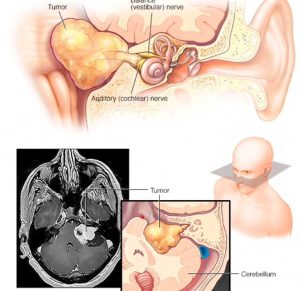
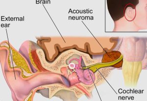
Acoustic neuromas, or vestibular schwannomas, are non-malignant fibrous tumors originating in the inner ear and auditory nerves. They do not spread to other parts of the body and they comprise from 6% to 10% of all brain tumors.
What are the symptoms of acoustic neuroma?
Acoustic neuromas are located in deep regions of the brain close to vital structures. Initial symptoms are hearing and balance problems. As a neuroma grows, the likelihood of compromising even more vital nerves increases, and new symptoms arise such as headaches. This can be the result of increased intracranial pressure and, if there is significant tumor growth for long periods, this can be fatal.

How is acoustic neuroma diagnosed?
If a routine hearing test demonstrates hearing loss and impaired sound discrimination, a BERA or BAEP test (brainstem auditory evoked potential) can be performed. This test provides data about the transmission of sound information from the ear to the brain, and results may indicate that the acoustic nerve is not functioning properly. If there is an abnormality in the ABR test, a detailed imaging exam such as high-resolution computed tomography (HRCT) or nuclear magnetic resonance exam (NMR) is usually ordered.
Computed tomography takes a series of x-ray images from different angles and uses software to combine them and provide a detailed view into the body’s interior structures. This is not painful, however, sometimes a patient may feel discomfort inside the enclosed space of the equipment. Tomography can be effective in locating acoustic neuromas, but small tumors within the auditory canal may not appear in conventional tomography. In order to improve the exam´s performance, it may be necessary to inject the patient with a contrast substance before the exam in order to improve the detail in the resulting images.
Nuclear magnetic resonance (NMR) is a radiological technique that combines magnets and advanced computer technology to produce images of internal body structures. These provide very detailed images that allow for the detection of small alterations within the body. In the case of a possible acoustic neuroma, gadolinium contrast agent is used to improve the images and highlight the tumor. NMR is also painless and has the advantage of not exposing the patient to radiation in the way that tomography does.

What are the forms of acoustic neuroma?
Acoustic neuromas present in two ways, either sporadic or associated with neurofibromatosis type 2 (NF2). Up to 95% of neuromas are sporadic and unilateral, meaning that they affect only one ear. Generally, patients with sporadic forms of acoustic neuroma encounter their first symptoms in middle age, usually at around 50.
On the other hand, tumors associated with NF2 are bilateral, meaning that they affect both ears, and make up approximately 5% of acoustic neuroma cases. NF2 is a rare genetic condition that affects one in every 100,000 people in the US. These patients develop benign tumors on their auditory nerves, and symptoms appear at younger ages, on average at around 30.
What treatment is there for acoustic neuroma?
The translabyrinthine procedure involves an incision behind the ear, and the mastoid bone and inner ear are removed to expose the tumor. Usually, the acoustic neuroma is completely removed, whereas partial removal is performed in rare cases. The opening in the mastoid is usually closed with fat removed from the abdomen. This translabyrinthine procedure compromises hearing and the balance mechanism of the inner ear, and it leads to permanent hearing loss. If the balancing mechanism is removed from one ear, its partner in the other ear compensates, and stability returns in 1 to 4 months after the operation.
The middle fossa procedure involves an incision above the ear, and then the brain is elevated to expose the acoustic neuroma. The tumor is completely removed in most cases. This surgical technique often preserves hearing in the ear that tis operated on, and complete resection of the acoustic neuroma is quite common. In approximately 40% of cases, the tumor involves the auditory nerve and the artery supplying the inner ear, in which case resection of the acoustic neuroma will compromise the hearing in the affected ear.
The retrosigmoid procedure begins with as incision behind the ear and then the brain is compressed to expose the acoustic neuroma. Every effort is made to preserve hearing, however, in some cases, it is necessary to remove the auditory nerve to achieve total resection of the neuroma. In approximately 50% of cases, the acoustic neuroma impacts the auditory nerve and the artery that supplies the inner ear, and so in these cases tumor resection compromises the hearing in the affected ear. Some patients report a persistent headache after this procedure.
Is it possible to preserve hearing in the ear with acoustic neuroma?
The choice of treatment for acoustic neuroma depends on the size of the neuroma and the level of residual hearing, and in some cases it is possible to preserve residual hearing. If hearing preservation is achieved in the affected ear, at best, hearing can be maintained at the same preoperative level. In the case of a tumor that already presents with advanced hearing impairment prior to surgery, undertaking a procedure that result in complete hearing loss in the affected ear is indicated in order to safely remove the acoustic neuroma and protect the patient´s life.

